Recently, a research team from Stanford University published an article in Nature Biotechnology titled "Inferring gene expression from cell-free DNA fragmentation profiles." Building on the analysis of multi-gene expression levels from cfDNA sequencing, the study established a new method for analyzing cfDNA fragmentation patterns — "Epigenetic Expression Inference from Cell-free DNA-sequencing (EPIC-seq)" — which can be applied at the same time to early cancer detection, subtyping and companion diagnostics.
Background
Cell-free DNA (cfDNA) consists of degraded DNA fragments released into plasma. It is present in various body fluids, and its concentration changes in response to tissue injury, cancer and inflammatory reactions. In healthy individuals, cfDNA consists mainly of small, uniform fragments of 185–200 bp produced during apoptosis. Tumor necrotic tissue cells, undergoing an abnormal apoptotic process, generate larger fragments of varying size above 200 bp. In 1947, Mandel and Metais first reported the presence of cfDNA in peripheral blood. Under physiological conditions, cfDNA in blood derives mainly from the necrosis and apoptosis of leukocytes. In certain diseases and special states, other cells also release free DNA into the blood, including circulating tumor DNA (ctDNA) derived from tumor cells and cfDNA of fetal origin.
Detection of circulating tumor DNA (ctDNA) in blood brings hope for non-invasive cancer testing. Chromatin fragmentation features have been used to infer the gene expression profiles of cell-free DNA (cfDNA), but current fragmentation-based methods require high concentrations of tumor-derived DNA and offer limited resolution.
Methods
The researchers described promoter fragmentation as an epigenomic cfDNA feature that can predict the RNA expression level of an individual gene: targeted sequencing of a target gene's promoter allows the epigenetic expression level to be inferred. Analyzing 329 blood samples in total from 201 cancer patients and 87 healthy adults, they demonstrated subtyping of lung cancer and diffuse large B-cell lymphoma. In addition, the researchers applied EPIC-seq to serial blood samples collected at multiple time points from patients receiving PD-(L)1 immunotherapy and found that the inferred gene expression profiles were closely associated with the patients' clinical presentation. Together, this work shows that EPIC-seq enables non-invasive, high-throughput analysis of tumor tissue of origin, holding great potential in diagnosis, prognosis and treatment.
Results
Association between cfDNA molecular features and gene expression levels
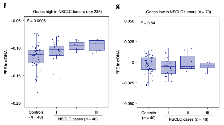
The researchers hypothesized that cfDNA fragments governed by active promoters are more likely to display random cleavage patterns than those governed by inactive promoters, making it possible to infer the expression of individual genes from cfDNA and to reflect the typing of various cell types from different tissue origins, including solid tumors. Through deep whole-genome sequencing analysis of samples from cancer patients of unknown origin, they found that cfDNA molecules mapped to highly expressed genes showed greater fragment length diversity than those mapped to lowly expressed genes. Comparing lung cancer patients with healthy controls, the researchers further established that promoter fragment entropy is significantly correlated with gene expression levels in non-small cell lung cancer (NSCLC). Genes highly expressed in NSCLC showed markedly elevated promoter fragment entropy in patient blood, and this was associated with lung cancer stage, whereas no such trend was seen for lowly expressed genes — indicating that this cfDNA feature is both gene- and tissue-specific.
Validating cfDNA-inferred gene expression levels in solid tumors
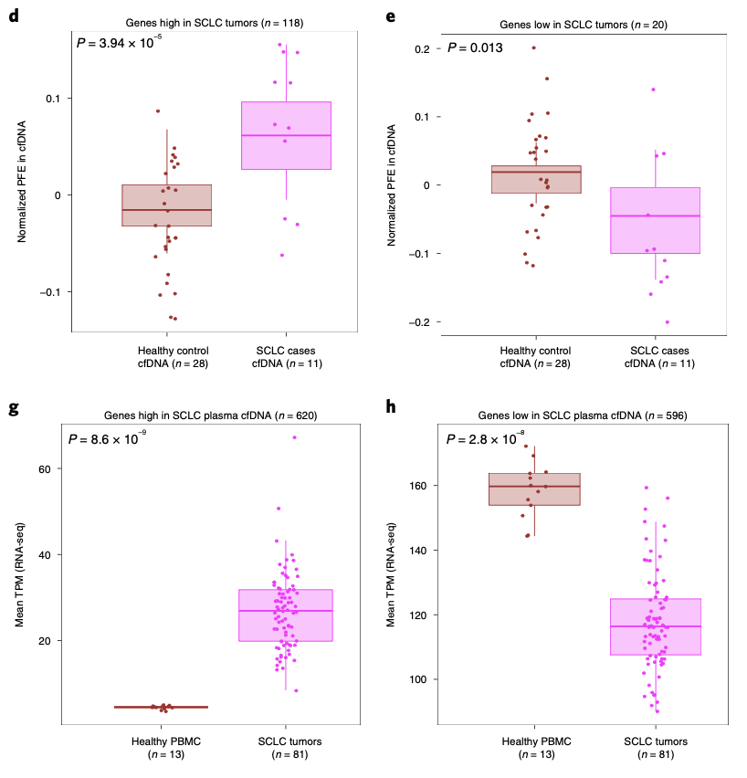
The researchers further tested whether whole-exome sequencing could confirm the accuracy of cfDNA-inferred expression levels. Performing ultra-deep whole-exome sequencing of plasma cfDNA from 11 patients with small cell lung cancer (SCLC) and 28 healthy controls, the authors found that for tumor genes highly expressed in primary SCLC tumors, promoter fragment entropy in patient plasma cfDNA was significantly higher than in healthy controls, while genes lowly expressed in SCLC showed the opposite trend. Combining these two sets of feature genes, the researchers derived a computational model that effectively distinguishes SCLC patients from healthy controls (AUC=0.98). Among these feature genes, they further screened and identified three SCLC signature marker genes: ASCL1, ANK1 and ASTN1.
Inferring gene expression from cfDNA fragmentation profiles (EPIC-seq)
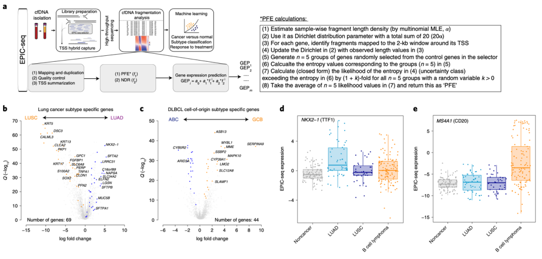
Greater sequencing depth improves the accuracy of cfDNA-inferred gene expression, so the authors developed EPIC-seq, which combines targeted deep sequencing of the TSS-flanking regions of cfDNA with machine learning to predict gene expression levels precisely. To validate the concept, the researchers applied EPIC-seq to the non-invasive histological subtyping of solid tumors and the molecular subtyping of hematological malignancies. First, they identified genes that are highly expressed in tumor tissue but relatively lowly expressed in whole blood, then compared differential expression between lung adenocarcinoma (LUAD) and lung squamous cell carcinoma (LUSC), and between GCB-type and ABC-type diffuse large B-cell lymphoma (DLBCL), thereby narrowing the field to tumor subtype-specific genes. Using the EPIC-seq workflow, the researchers analyzed cfDNA from 373 plasma samples from 288 adults, including 109 plasma samples from 87 NSCLC patients, 126 plasma samples from 114 DLBCL patients and 94 plasma samples from 87 healthy controls. Ultimately they identified NKX2-1, a gene highly expressed in LUAD, as an effective marker for lung cancer tissue typing, and MS4A1 (CD20), highly expressed in DLBCL, as an effective marker for immunophenotypic subtyping of lymphoma.
EPIC-seq for the non-invasive detection and subtyping of lung cancer and for predicting response to PD-(L)1 immunotherapy
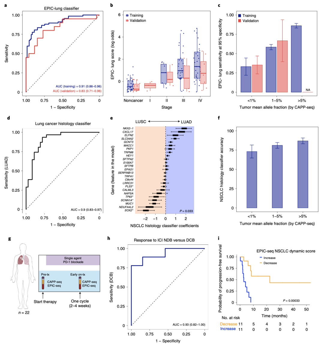
Using machine learning, the researchers built a model comparing lung cancer patients with healthy controls, randomly dividing the cohort into a training set and a validation set. The training set contained 67 lung cancer patients (7 stage II, 30 stage III, 30 stage IV) and 71 healthy controls; the validation set contained 20 lung cancer patients and 23 healthy controls. AUC values in the training and validation sets were 0.91 and 0.83 respectively, showing good lung cancer detection performance, and EPIC scores for patients with stage III–IV disease were higher than for stage II. To examine the use of EPIC-seq in lung cancer subtyping, the researchers selected a cohort of 67 lung cancer patients, divided by histological type into a lung adenocarcinoma group (36 cases) and a lung squamous carcinoma group (31 cases), and achieved good subtyping performance (AUC=0.90) using training and cross-validation. For patients with advanced NSCLC, PD-(L)1 immunotherapy is critically important, yet its efficacy and toxic side effects remain poorly defined, and reliable biomarkers that accurately predict whether a patient will benefit before or early in treatment are lacking. The researchers therefore used EPIC-seq to analyze blood samples from 22 NSCLC patients receiving PD-(L)1 immunotherapy, collected before treatment and in the four weeks prior to the start of treatment, and developed a "lung dynamic factor" that effectively predicts treatment response.
EPIC-seq for the non-invasive detection and molecular subtyping of diffuse large B-cell lymphoma (DLBCL)
DLBCL is a common non-Hodgkin lymphoma with marked clinical and biological heterogeneity. Clinically, this heterogeneity can be captured through the International Prognostic Index (IPI) and genotyping of the primary tumor biopsy, but effective, minimally invasive detection methods are still lacking. The researchers applied EPIC-seq to analyze blood cfDNA from 91 DLBCL patients and 71 healthy controls, training a discrimination model that performed well (AUC=0.92); in the validation set (23 DLBCL patients and 23 healthy controls) the AUC reached 0.96, and EPIC analysis showed a significant grading correlation with IPI. Further analysis of data from DLBCL patients before and after treatment confirmed that EPIC-seq can detect tissue-derived DLBCL signals non-invasively and faithfully reflect clinical presentation before and after therapy. DLBCL is divided into two subtypes by cell of origin; patients with the GCB subtype generally have a better prognosis and may show greater sensitivity to targeted therapy. The researchers therefore carried out further EPIC-seq analysis of pre-treatment samples and derived a model score that distinguishes the GCB subtype well, correlating strongly with subtyping results obtained at the tumor tissue level (r=0.84).
Summary and outlook
The researchers introduced a method for non-invasive inference of gene expression using cfDNA fragmentation patterns — EPIC-seq — which can be applied widely in clinically relevant settings including tumor detection, subtype classification, response assessment and prognostic analysis. Further refinement is still needed to make the most of this fragmentation-based technology. Patients with localized tumors show considerable molecular heterogeneity, and low ctDNA levels may prevent its direct use in early cancer detection. As more genes and more fragmentomic features are investigated with this approach, stronger signals can be obtained for the non-invasive detection of small tumor deposits, enabling more specific study of signaling pathway activity and tissue pathology.
References:
Mohammad Shahrokh Esfahani, Emily G. Hamilton et al. Inferring gene expression from cell-free DNA fragmentation profiles. Nature Biotechnology, 2022, 40: 585-597.